| Glycophorin A | |||||||||
|---|---|---|---|---|---|---|---|---|---|
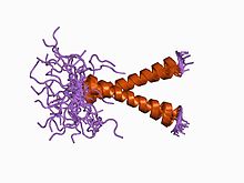
 Dimeric transmembrane domain of human glycophorin A (20 NMR-determined structures) | |||||||||
| Identifiers | |||||||||
| Symbol | GYPA | ||||||||
| Pfam | PF01102 | ||||||||
| InterPro | IPR001195 | ||||||||
| PROSITE | PDOC00281 | ||||||||
| SCOP2 | 1afo / SCOPe / SUPFAM | ||||||||
| OPM superfamily | 25 | ||||||||
| OPM protein | 5eh4 | ||||||||
| Membranome | 156 | ||||||||
| |||||||||
A glycophorin is a sialoglycoprotein of the membrane of a red blood cell. It is a membrane-spanning protein and carries sugar molecules. It is heavily glycosylated (60%). Glycophorins are rich in sialic acid, which gives the red blood cells a very hydrophilic-charged coat. This enables them to circulate without adhering to other cells or vessel walls.
A particular mutation in Glycophorins is thought to produce a 40% reduction in risk of severe malaria. [1]
Identification
After separation of red cell membranes by SDS-polyacrylamide gel electrophoresis and staining with periodic acid-Schiff staining (PAS), four glycophorins have been identified. These have been named glycophorin A, B, C, and D in order of the quantity present in the membrane, glycophorin A being the most and glycophorin D the least common. A fifth ( glycophorin E) has been identified within the human genome but cannot easily be detected on routine gel staining. In total, the glycophorins constitute ~2% of the total erythrocyte membrane protein mass. These proteins are also known under different nomenclatures but they are probably best known as the glycophorins.
Family members
The following four human genes encode glycophorin proteins:
Glycophorin D is now known to be a variant of Glycophorin C.
References
- ^ Leffler EM, Band G, Busby GB, Kivinen K, Le QS, Clarke GM, et al. (June 2017). "Resistance to malaria through structural variation of red blood cell invasion receptors". Science. 356 (6343): eaam6393. doi: 10.1126/science.aam6393. PMC 5575826. PMID 28522690.
External links
- Glycophorin at the U.S. National Library of Medicine Medical Subject Headings (MeSH)
- UMich Orientation of Proteins in Membranes protein/pdbid-1afo