|
| Warning This page contains syntax errors ("cite%20note") caused by a VisualEditor bug. Do not copy/move content from this page until the errors have been repaired. See {{
Warning VisualEditor bug}} for more information. |
 | This is a user sandbox of
Ryansnow. You can use it for testing or practicing edits. This is not the sandbox where you should draft your assigned article for a dashboard.wikiedu.org course. To find the right sandbox for your assignment, visit your Dashboard course page and follow the Sandbox Draft link for your assigned article in the My Articles section. |
(Pacific Lutheran University Comparative Anatomy dissection)
Week 13
5/3/2019
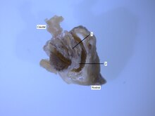
Re-highlighted the Agistrodon mokasen figure in powerpoint and downloaded the microscope photo from lab.
ORIGINAL
Pit vipers have a specialized sensory organs near the nostrils called heat sensing pits. [1] The location of this organ is unique to pit vipers. These pits have the ability to detect thermal radiation emitted by warm-blooded animals which helps them better understand their environment. [2] Internally the organ forms a small pit lined with membranes attached to the trigeminal nerve. [1] [3] Infrared light signals the internal membranes and the trigeminal nerves send the infrared signals to the brain where they are overlaid onto the visual image created by the eyes. [4]
EDITED
Pit vipers have specialized sensory organs near the nostrils called heat sensing pits. [3] The location of this organ is unique to pit vipers. These pits have the ability to detect thermal radiation emitted by warm-blooded animals, helping them better understand their environment. [1] Internally the organ forms a small pit lined with membranes, external and internal, attached to the trigeminal nerve. [3] [5]Infrared light signals the internal membranes, which in turn signal the trigeminal nerve and send the infrared signals to the brain where they are overlaid onto the visual image created by the eyes. [4]
Week 12
Moving Work 4/22/2019
Images from the dissection into the rattlesnake page


Week 10
Second Draft 4/12/2019
Original Article
Aside from this pair of simple eyes, rattlesnakes are able to detect thermal radiation emitted by warm-blooded organisms in their environment. Functioning optically like a pinhole camera eye, thermal radiation, in the form of infrared wavelength light, enters, passes through the opening of the pit and strikes the pit membrane located in the back wall, warming this part of the organ. Due to the extremely high density of these heat-sensitive receptors innervating this membrane, the rattlesnake can detect temperature changes of 0.003 °C or less in its immediate surroundings. Infrared cues from these receptors are transmitted to the brain by the trigeminal nerve, where they are used to create thermal maps of the snake’s surroundings. Due to the small sizes of the pit openings, typically these thermals images are low in resolution and contrast. Nevertheless, rattlesnakes superimpose visual images created from information from the eyes with these thermal images from the pit organs to more accurately visualize their surroundings in low levels of light. Research conducted recently on the molecular mechanism of this ability suggests the temperature sensitivity of these pit organs is closely linked to the activity of transient receptor potential ankyrin 1, a temperature-sensitive ion channel saturated in the pit membrane.
With Citations and Links
Aside from this pair of simple eyes, rattlesnakes are able to detect thermal radiation emitted by warm-blooded organisms in their environment. [2] Functioning optically like a pinhole camera eye, thermal radiation, in the form of infrared wavelength light, enters, passes through the opening of the pit and strikes the pit membrane located in the back wall, warming this part of the organ. [5] [1] Due to the extremely high density of these heat-sensitive receptors innervating this membrane, the rattlesnake can detect temperature changes of 0.003 °C or less in its immediate surroundings. [5] Infrared cues from these receptors are transmitted to the brain by the trigeminal nerve, where they are used to create thermal maps of the snake’s surroundings. [4] Due to the small sizes of the pit openings, typically these thermals images are low in resolution and contrast. Nevertheless, rattlesnakes superimpose visual images created from information from the eyes with these thermal images from the pit organs to more accurately visualize their surroundings in low levels of light. [4] Research conducted recently on the molecular mechanism of this ability suggests the temperature sensitivity of these pit organs is closely linked to the activity of transient receptor potential ankyrin 1, a temperature-sensitive ion channel saturated in the pit membrane.
For
Viperidae
Pit vipers have a specialized sensory organs near the nostrils called heat sensing pits. [1] The location of this organ is unique to pit vipers. These pits have the ability to detect thermal radiation emitted by warm-blooded animals which helps them better understand their environment. [2] Internally the organ forms a small pit lined with membranes attached to the trigeminal nerve. [1] [3] Infrared light signals the internal membranes and the trigeminal nerves send the infrared signals to the brain where they are overlaid onto the visual image created by the eyes. [4]
Week 9
Peer Review 4/8/2019
"The inclusion of actual dissection images will be great to avoid copyright issues and directly represent the topics you guys have chosen"
- I plan on exposing the internal membranes and hopefully the trigeminal nerve during our dissection so that I can label those parts on a real specimen.
To address comments from Dr. Schutz on my first draft: I am going to continue adding citations to the heat sensing pit section on the rattlesnake page but add a a section on the Viperidae page because they have a particular type of infrared pit. I plan on going into detail about the trigeminal nerve because it plays such an important role in the sensing process. In addition, I am also looking for images from old (pre 1944) papers for a diagram of the infrared pit.

Week 6
Draft 3/15/2019
CHANGES: Heat sensing pits are organs on a rattlesnakes’ face that detect infrared heat from warm-blooded organisms. [2] The pit is located between the nostril and the eye on both sides of the face. The interior of the pit has a membrane on the back wall that is lined with clusters of nerve endings that transmit the infrared signals to the brain. [5] Rattlesnakes are extremely sensitive to temperatures and can detect 0.003°C changes in temperatures making it possible for them to differentiate the environment and prey. [5] Once the infrared signals are detected, the nerve clusters send this information to the brain via the trigeminal nerve. Once the signal reaches the brain, it is superimposed onto the visual image the snake is also receiving allowing them to hunt or escape predators more efficiently. [4]
ORIGINAL ARTICLE: Aside from this pair of simple eyes, rattlesnakes are able to detect thermal radiation emitted by warm-blooded organisms in their environment. Functioning optically like a pinhole camera eye, thermal radiation, in the form of infrared wavelength light, enters, passes through the opening of the pit and strikes the pit membrane located in the back wall, warming this part of the organ. Due to the extremely high density of these heat-sensitive receptors innervating this membrane, the rattlesnake can detect temperature changes of 0.003 °C or less in its immediate surroundings. Infrared cues from these receptors are transmitted to the brainby the trigeminal nerve, where they are used to create thermal maps of the snake’s surroundings. Due to the small sizes of the pit openings, typically these thermals images are low in resolution and contrast. Nevertheless, rattlesnakes superimpose visual images created from information from the eyes with these thermal images from the pit organs to more accurately visualize their surroundings in low levels of light. Research conducted recently on the molecular mechanism of this ability suggests the temperature sensitivity of these pit organs is closely linked to the activity of transient receptor potential ankyrin 1, a temperature-sensitive ion channel saturated in the pit membrane.
Week 5
Find Your Sources 3/8/2019
I was chosen for the rattlesnake dissection team. I will be posting this in the talk page of the rattlesnake page to see if any other users have ideas:
The Heat Sensing Pit section has no referances along with an uninformative picture. I would like to update this section by double checking the information and adding sources to the paragraph and using a different photo.
Sources:
"Biological Infrared Imaging and Sensing [2]
The Infrared "Vision" of Snakes [5]
Properties of an Infrared Receptor [3]
Molecular Basis of Infrared Detection by Snakes [4]
Week 4
Project Prefernces 3/1/2019
1. Mudpuppy: Last semester I wrote a paper on giant salamanders so it would be interesting to explore a new kind of salamander Pages that could be added to:
- Cloaca: Page lists that amphibians have cloacas but does not discuss it and the common mudpuppy page links it to this page.
-The Respriratory System: You can see there are external gills based on the photo but there is no mention of how they work
-Teeth are discussed at great length but homodont is linked to heterodont. It would probably be better not to link it at all
2. Rattlesnake: It would be interesting to see the internal anatomy of the snake because they have to make internal alterations due to their long bodies and occillating movement
-There is a lot of information on the heat sensing pits but there are no references and the photo doesn't do a good job at pointing them out.
- No skull morphology is mentioned or even linked to the snake skeleton page. The jaws are particularly interesting because they allow the snake to eat things much larger than its size
-The reproductive system could be improved. There is more information about male anatomy than female even though females give live birth
3. Rabbit: It would be interesting to see a herbivores digestive tract
- There are no pictures of the rabbit cecum and on the cecum page they only use human anatomy and cockroach photos to try and illustrate where the cecum is. Having an image of a mammal that actually has a cecum would be beneficial
- The hindgut page has very little information and all the images are of human embryos and worms. The rabbit page very clearly explains how hidgut digestion works but the actual hindgut digestion page does not
-Overall the digestion section needs citations
Week 3
Adding to an Article 2/22/2019
Lamprey Article Edits
ARTICLE: Adults superficially resemble eels in that they have scaleless, elongated bodies, and can range from 13 to 100 cm (5 to 40 inches) in length. Lacking paired fins, adult lampreys have large eyes, one nostril on the top of the head, and seven gill pores on each side of the head. As part of courtship, male sea lampreys rub a ridge of back tissue against females’ abdomens. [6]
ARTICLE: Research on sea lampreys has revealed that sexually mature males use a specialized heat-producing tissue in the form of a ridge of fat cells near the anterior dorsal fin to stimulate females. After having attracted a female with pheromones, the heat detected by the female through body contact will encourage spawning. [7]
CHANGE: The last sentence of the first paragraph doesn't belong there and it has the same information as the second paragraph. I am going to delete the sentence from the first paragraph.
ARTICLE: Near the gills are the eyes, which are poorly developed and buried under skin in the larvae. The eyes complete their development during metamorphosis, and are covered by a thin and transparent layer of skin that becomes opaque in preservatives. [8] Their teeth consist of a meshwork of keratin filaments and other proteins. [9]
ARTICLE: The last common ancestor of lampreys appears to have been specialized to feed on the blood and body fluids of other fish after metamorphosis. They attach their mouthparts to the target animal's body, then use three horny plates (laminae) on the tip of their piston-like tongue, one transversely and two longitudinally placed, to scrape through surface tissues until they reach body fluids. The teeth on their oral disc are primary used to help the animal attach itself to its prey. The teeth have a hollow core to give room for replacement teeth growing under the old ones. Some of the original blood-feeding forms have evolved into species that feed on both blood and flesh, and some who have become specialized to eat flesh and may even invade the internal organs of the host. Tissue feeders can also involve the teeth on the oral disc in the excision of tissue. [10] [11] [12] [13] [14] [15]
CHANGE: The last sentence of the first paragraph has very similar wording as the source the information came from and has nothing to do with this paragraph so I will move it to the paragraph about feeding by adding the information to an existing sentence.
NEW SENTENCE: Made of keratin and other proteins, lamprey teeth have a hollow core to give room for replacement teeth growing under the old ones
Week 2
Article Review 2/15/2019
I think that the first paragraph could be written more clearly/sound better. It reads like the author didn't proof read before publishing. There are certain areas where mating displays are mentioned out of context and could simply be put together in the Lifecycle section. I couldn't find any areas of bias. Many of the sources were journal articles and all of the links I chose to look at were functioning and would direct you to the reference. The references were correctly formatted with numbers at the ends of sentences. A source I examined discussed the keratinous teeth that lampreys have. Both in the article and the source the word "meshwork" was used to discuss the arrangement of the teeth. The talk page shows that the last time anyone discussed the lamprey page was 2017 but in the history edits were made in January 2019. Wikipedia isn't as scientific about the subject as were are class but that is because it is built for the general public.
References
- ^ a b c d e Lynn, W. Gardner (1931). "The Structure and Function of the Facial Pits of Pit Vipers". American Journal of Anatomy. 49: 97–139.
- ^ a b c d e Campbell, Angela L.; Naik, Rajesh R.; Sowards, Laura; Stone, Morley O. (2002). "Biological Infrared Imaging and Sensing" (PDF). Micron. 33: 211-225.
- ^ a b c Bullock, T.H.; Diecke, F.P.J. (1956). "Properties of an Infrared Receptor". Journal of Physiology. 134: 47-87.
- ^ a b c d e f Gracheva, Elena O.; Ingolia, Nicolas T.; Kelly, Yvonne M.; Cordero-Morales, Julio F.; Hollopeter, Gunther; Chesler, Alexander T.; Sanchez, Elda E.; Perez, John C.; Weissman, Johnathan S.; Julius, David (2010). "Molecular Basis of Infrared Detection by Snakes". Nature. 464 (7291): 1006-1011.
- ^ a b c d e Newman, Eric A.; Hartline, Peter H. (1982). "The Infrared "Vision" of Snakes". Scientific American. 246 (3): 116-127.
-
^
https://www.the-scientist.com/the-nutshell/lampreys-heat-up-before-mating-39114.
{{
cite journal}}
: Cite has empty unknown parameter:|1=( help); Cite journal requires|journal=( help); Missing or empty|title=( help) - ^ Poppick, Laura (2015-11-02). "Only the hot sea lamprey guys get sex -- thermally, that is". NBC News.
- ^ Iuliis, Gerardo De; Deiuliis, Gerald; Pulera, Dino (2006-08-03). The Dissection of Vertebrates: A Laboratory Manual. ISBN 9780080477350.
- ^ International Review of Cell and Molecular Biology. 2016-09-30. ISBN 9780128052204.
- ^ Rohde, Klaus (2005-09-13). Marine Parasitology. ISBN 9780643099272.
- ^ Ehrlich, Hermann (2014-12-01). Biological Materials of Marine Origin: Vertebrates. ISBN 9789400757301.
-
^ Warren, Melvin L., Jr; Burr, Brooks M. (2014-07-10).
Freshwater Fishes of North America: Volume 1: Petromyzontidae to Catostomidae.
ISBN
9781421412016.
{{
cite book}}
: CS1 maint: multiple names: authors list ( link) - ^ Khidir, K. Teresa (2003). "Oral fimbriae and papillae in parasitic lampreys (Petromyzontiformes)". Environmental Biology of Fishes. 66 (3): 271–278. doi: 10.1023/A:1023961910547.
- ^ Potter, Ian C.; Gill, Howard S. (2003). "Adaptive Radiation of Lampreys". Journal of Great Lakes Research. 29: 95–112. doi: 10.1016/S0380-1330(03)70480-8.
- ^ Barras, Colin (2 November 2015). "Meet a lamprey. Your ancestors looked just like it". BBC Earth.