| Tunica albuginea of testis | |
|---|---|
 A diagram of the major components of an adult human testicle, including the following numbered items: 1. Tunica albuginea, 2.
Septula testis, 3.
Lobulus testis, 4.
Mediastinum testis, 5.
Tubuli seminiferi contorti, 6.
Tubuli seminiferi recti, 7.
Rete testis, 8.
Ductuli efferentes testis, 9a. Head of
epididymis, 9b. Body of epididymis, 9c. Tail of epididymis, 10.
Vas deferens, 11a.
Tunica vaginalis (parietal lamina), 11b. Tunica vaginalis (visceral lamina), and 12.
Cavity of tunica vaginalis. | |
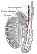
 Section of a testicle of a
bull, blood vessels injected with red gelatine. 1
parenchyma, 2
mediastinum testis, 3
tunica albuginea, 4 tail of
epididymis, 5 head of epididymis, 6
spermatic cord with
convoluted testicular artery | |
| Details | |
| Identifiers | |
| Latin | tunica albuginea testis |
| FMA | 19843 |
| Anatomical terminology | |
The tunica albugine is a dense, [1] [2] blue-white [3] layer of fibrous tissue surrounding the testis. [1] [4] It is the middle of three envelopes forming the capsule of the testis; it is deep to the visceral layer of tunica vaginalis, and superficial to the tunica vasculosa testis (vascular layer of testis). [5]
The connective tissue of the tunica albuginea testis extends into the substance of the testis to form fibrous partitions - the septa testis. [1] At the posterior aspect of the testis (where the serosa of testis is deficient to allow for the attachment of the epididymis), the tunica albuginea extends into the testis to form the mediastinum testis. [5]
Anatomy
It is thicker than the tunica albuginea of the ovary. [6]
Histology
It is composed of bundles of white fibrous connective tissue (from which it derives its name albuginea) which interlace in every direction. [7]
Additional images
-
 Transverse section through the left side of the scrotum and the left testis.
 -
 Section of a genital cord of the testis of a human embryo 3.5 cm. long.
 -
 Vertical section of the testis, to show the arrangement of the ducts.


References
![]() This article incorporates text in the
public domain from
page 1242 of the 20th edition of
Gray's Anatomy (1918)
This article incorporates text in the
public domain from
page 1242 of the 20th edition of
Gray's Anatomy (1918)
- ^ a b c Martini, Frederic; Tallitsch, Robert B.; Nath, Judi L. (2017). Human Anatomy (9th ed.). Pearson. p. 711. ISBN 9780134320762.
-
^ Standring, Susan (2020).
Gray's Anatomy: The Anatomical Basis of Clinical Practice (42nd ed.). New York. p. 1292.
ISBN
978-0-7020-7707-4.
OCLC
1201341621.
{{
cite book}}
: CS1 maint: location missing publisher ( link) -
^ Standring, Susan (2020).
Gray's Anatomy: The Anatomical Basis of Clinical Practice (42nd ed.). New York. p. 1292.
ISBN
978-0-7020-7707-4.
OCLC
1201341621.
{{
cite book}}
: CS1 maint: location missing publisher ( link) - ^ Federle, Michael P.; Rosado-de-Christenson, Melissa L.; Raman, Siva P.; Carter, Brett W., eds. (2017-01-01), "Testes and Scrotum", Imaging Anatomy: Chest, Abdomen, Pelvis (Second Edition), Elsevier, pp. 1000–1017, doi: 10.1016/B978-0-323-47781-9.50043-X, ISBN 978-0-323-47781-9, retrieved 2021-02-03
- ^
a
b Standring, Susan (2020).
Gray's Anatomy: The Anatomical Basis of Clinical Practice (42nd ed.). New York. p. 1292.
ISBN
978-0-7020-7707-4.
OCLC
1201341621.
{{
cite book}}
: CS1 maint: location missing publisher ( link) - ^ Hummitzsch, Katja; Irving-Rodgers, Helen F.; Schwartz, Jeff; Rodgers, Raymond J. (2019-01-01), Leung, Peter C. K.; Adashi, Eli Y. (eds.), "Chapter 4 - Development of the Mammalian Ovary and Follicles", The Ovary (Third Edition), Academic Press, pp. 71–82, ISBN 978-0-12-813209-8, retrieved 2021-02-03
- ^ Gray, Henry; Lewis, Warren Harmon (1918). Anatomy of the human body. Harold B. Lee Library (42nd ed.). Philadelphia : Lea & Febiger. p. 1242.
External links
- Anatomy photo:36:11-0102 at the SUNY Downstate Medical Center - "Inguinal Region, Scrotum and Testes: The Cross-Section of the Testis"
- inguinalregion at The Anatomy Lesson by Wesley Norman (Georgetown University) ( testes)
|
| This article related to the genitourinary system is a stub. You can help Wikipedia by expanding it. |