| Superior epigastric artery | |
|---|---|
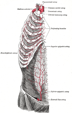
 Superior epigastric artery,
internal thoracic artery and
inferior epigastric artery. (Superior epigastric artery is labeled at right center.) | |
| Details | |
| Source | internal thoracic |
| Vein | superior epigastric vein |
| Identifiers | |
| Latin | arteria epigastrica superior |
| TA98 | A12.2.08.041 |
| TA2 | 4588 |
| FMA | 10646 |
| Anatomical terminology | |
In human anatomy, the superior epigastric artery is a terminal [1] branch of the internal thoracic artery that provides arterial supply to the abdominal wall, and upper rectus abdominis muscle. It enters the rectus sheath to descend upon the inner surface of the rectus abdominis muscle. It ends by anastomosing with the inferior epigastric artery.
Structure
Origin
The superior epigastric artery arises from the internal thoracic artery (referred to as the internal mammary artery in the accompanying diagram). [2] [3]
Course and relations
The superior epigastric artery pierces the diaphragm [1] to enter the rectus sheath and descend upon the deep surface of the rectus abdominis. [4]
Along its course, it is accompanied by a similarly named vein, the superior epigastric vein.[ citation needed]
Anastomoses
It anastomoses with the inferior epigastric artery [4] [2] within the rectus abdominis muscle [4] [1] at the umbilicus. [2]
Distribution
Where it anastomoses, the superior epigastric artery supplies the anterior part of the abdominal wall, [5] [6] upper rectus abdominis muscle, [5] and some of the diaphragm.[ citation needed]
Collateralization in disease
Vascular disease
The superior epigastric arteries, inferior epigastric arteries, internal thoracic arteries and left subclavian artery and right subclavian artery / brachiocephalic are collateral vessels to the thoracic aorta and abdominal aorta. If the abdominal aorta develops a significant stenosis and/or blockage (as may be caused by atherosclerosis), this collateral pathway may develop sufficiently, over time, to supply blood to the lower limbs. [7]
Coarctation of the aorta
A congenitally narrowed aorta, due to coarctation, is often associated with a significant enlargement of the internal thoracic and epigastric arteries. [8]
See also
References
- ^ a b c Sinnatamby, Chummy (2011). Last's Anatomy (12th ed.). Elsevier Australia. p. 225. ISBN 978-0-7295-3752-0.
- ^ a b c Castro Ferreira, Marcus; Henrique Ishida, Luis; Munhoz, Alexandre (January 1, 2009), Wei, Fu-Chan; Mardini, Samir (eds.), "CHAPTER 19 - Rectus flap", Flaps and Reconstructive Surgery, Edinburgh: W.B. Saunders, pp. 207–223, doi: 10.1016/b978-0-7216-0519-7.00019-8, ISBN 978-0-7216-0519-7, retrieved November 22, 2020
- ^ Ahmed, Abdul (January 1, 2017), Brennan, Peter A.; Schliephake, Henning; Ghali, G. E.; Cascarini, Luke (eds.), "37 - Common Free Vascularized Flaps: The Rectus Abdominis", Maxillofacial Surgery (Third Edition), Churchill Livingstone, pp. 533–542, doi: 10.1016/b978-0-7020-6056-4.00038-1, ISBN 978-0-7020-6056-4, retrieved November 22, 2020
- ^
a
b
c
The Big Picture: Gross Anatomy, Medical Course & Step 1 Review. David A. Morton, K. Bo Foreman, Kurt H. Albertine (2nd ed.). New York. 2018.
ISBN
978-1-259-86264-9.
OCLC
1044772257.
{{
cite book}}
: CS1 maint: location missing publisher ( link) CS1 maint: others ( link) - ^ a b Shell, Dan H.; Vásconez, Luis O.; de la Torre, Jorge I.; Chin, Gloria; Weinzweig, Norman (January 1, 2010), Weinzweig, Jeffrey (ed.), "Chapter 91 - Abdominal Wall Reconstruction", Plastic Surgery Secrets Plus (Second Edition), Philadelphia: Mosby, pp. 594–604, doi: 10.1016/b978-0-323-03470-8.00091-0, ISBN 978-0-323-03470-8, retrieved November 22, 2020
- ^ DiEdwardo, Christine A.; Caterson, Stephanie A.; Barrall, David T. (January 1, 2010), Weinzweig, Jeffrey (ed.), "Chapter 80 - Abdominoplasty", Plastic Surgery Secrets Plus (Second Edition), Philadelphia: Mosby, pp. 532–537, doi: 10.1016/b978-0-323-03470-8.00080-6, ISBN 978-0-323-03470-8, retrieved November 22, 2020
- ^ Yurdakul M, Tola M, Ozdemir E, Bayazit M, Cumhur T (April 2006). "Internal thoracic artery-inferior epigastric artery as a collateral pathway in aortoiliac occlusive disease". J. Vasc. Surg. 43 (4): 707–13. doi: 10.1016/j.jvs.2005.12.042. PMID 16616225.
- ^ Huhmann W, Kunitsch G, Dalichau H (1976). "[Coarctation of the aorta on the plain chest x-ray (author's transl)]". Dtsch Med Wochenschr. 101 (41): 1477–81. doi: 10.1055/s-0028-1104294. PMID 964150. S2CID 260093972.
External links
- Anatomy photo:18:07-0103 at the SUNY Downstate Medical Center - " Thoracic wall: Branches of the Internal Thoracic Artery"
- Anatomy figure: 35:04-03 at Human Anatomy Online, SUNY Downstate Medical Center - "Incisions and the contents of the rectus sheath. "