| Pituicyte | |
|---|---|
| Details | |
| Location | Posterior pituitary |
| Function | Glial cell |
| Identifiers | |
| Latin | pituicytus |
| NeuroLex ID | sao1004082033 |
| TH | H3.08.02.2.00040 |
| FMA | 83503 |
| Anatomical terms of microanatomy | |
Pituicytes are glial cells of the posterior pituitary. Their main role is to assist in the storage and release of neurohypophysial hormones. [1]
Structure
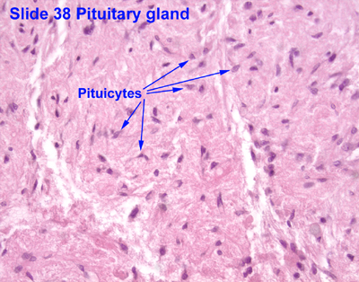
Pituicytes are located in the pars nervosa of the posterior pituitary and interspersed with unmyelinated axons and Herring bodies. They generally stain dark purple with an H&E stain and are among the easiest structures to identify in the region. [2] [3] Pituicytes have an irregular and branched shape which resembles that of another type of glial cell: the astrocyte. [4] Like astrocytes, their cytoplasm presents specific intermediate filaments made up of glial fibrillary acidic protein (GFAP). [5]
Function
Pituicytes are similar to astrocytes, another type of glial cell. Their main role is to assist in the storage and release of hormones of the posterior pituitary. Pituicytes surround axonal endings and regulate hormone secretion by releasing their processes from these endings. [1]
Clinical significance
Pituicytomas are rare tumors that arise from pituicytes. They may be mistaken for the much more common pituitary adenoma, as well as craniopharyngioma and meningioma. Symptoms from the mass effect of the tumor usually include vision disorders, and less often headaches, hypopituitarism (decreased function of the pituitary gland), fatigue, and decreased libido. [6]
See also
List of distinct cell types in the adult human body
References
- ^ a b Hatton, GI (September 1988). "Pituicytes, glia and control of terminal secretion" (PDF). The Journal of Experimental Biology. 139: 67–79. PMID 3062122.
- ^ Histology image: 38_08 at the University of Oklahoma Health Sciences Center
- ^ Histology image:14003loa from Vaughan, Deborah (2002). A Learning System in Histology: CD-ROM and Guide. Oxford University Press. ISBN 978-0195151732.
- ^ Ross M, Pawlina W (2011). Histology: A Text and Atlas (6th ed.). Lippincott Williams & Wilkins. p. 751. ISBN 978-0-7817-7200-6.
- ^ Wei, XY; Zhao, CH; Liu, YY; Wang, YZ; Ju, G (2009). "Immuohistochemical markers for pituicyte". Neuroscience Letters. 465 (1): 27–30. doi: 10.1016/j.neulet.2009.06.059. PMID 19559073. S2CID 39871786.
- ^ Covington, MF; Chin, SS; Osborn, AG (December 2011). "Pituicytoma, spindle cell oncocytoma, and granular cell tumor: clarification and meta-analysis of the world literature since 1893". American Journal of Neuroradiology. 32 (11): 2067–2072. doi: 10.3174/ajnr.A2717. PMC 7964422. PMID 21960498.