| Pes anserinus | |
|---|---|
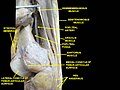
 Muscles of the gluteal and posterior femoral regions. Area of pes anserinus is encircled at bottom.
Sartorius,
gracilis,
semitendinosus and
semimembranosus are labeled at bottom left. | |
| Details | |
| Identifiers | |
| Latin | pes anserinus |
| TA2 | 2612 |
| FMA | 311256 |
| Anatomical terminology | |
Pes anserinus (" goose foot") refers to the conjoined tendons of three muscles of the thigh. Pes means 'foot' in Latin. In Latin, anser means 'goose', and anserinus means 'goose-like'.
Pes anserinus inserts onto the anteromedial (front and inside) surface of the proximal tibia. The muscles are the sartorius, gracilis and semitendinosus sometimes referred to as the guy ropes. The name "goose foot" arises from the three-pronged manner in which the conjoined tendon inserts onto the tibia.
Structure
The three tendons, from front to back, that conjoin to form the pes anserinus come from the sartorius muscle, the gracilis muscle, and the semitendinosus muscle. [1] [2] It inserts onto the proximal anteromedial surface of the tibia. [2]
The pes anserinus is around 5 cm below the medial tibial joint line. [2] It lies superficial to the tibial insertion of the medial collateral ligament of the knee. [1]
Clinical significance
Pes anserinus tendinitis/bursitis syndrome, or pes anserine bursitis, is a cause of chronic knee pain and weakness. [3] [4] It occurs when the medial portion of the knee is inflamed. If the bursa underlying the tendons of the sartorius, gracilis, and semitendinosus gets irritated from overuse or injury, a person can develop this ailment. This condition usually occurs in athletes from overuse. This pathology is characterized by pain, swelling, and / or tenderness. [3]
The semitendinosus tendon can be used in certain techniques for reconstruction of the anterior cruciate ligament. [5]
History
The name "goose foot" arises from the three-pronged manner in which the conjoined tendon inserts onto the tibia. [6]
Additional images
-
 Muscles of the posteromedial thigh, medial view.

References
- ^ a b Cai, Shaoren; Jin, Yong; Zhang, Wanyu; Liao, Zhongtang (2019-09-01). "Bloodletting-cupping for 169 cases of pes anserinus myotenositis". World Journal of Acupuncture - Moxibustion. 29 (3): 231–234. doi: 10.1016/j.wjam.2019.08.001. ISSN 1003-5257. S2CID 201960589.
- ^ a b c Finnoff, Jonathan T.; Nutz, David J.; Henning, Philip T.; Hollman, John H.; Smith, Jay (2010). "Accuracy of Ultrasound-Guided versus Unguided Pes Anserinus Bursa Injections". PM&R. 2 (8): 732–739. doi: 10.1016/j.pmrj.2010.03.014. ISSN 1934-1563. PMID 20598959. S2CID 26845656.
- ^ a b pmr/104 at eMedicine - "Pes anserinus bursitis"
- ^ Alvarez-Nemegyei J (April 2007). "Risk factors for pes anserinus tendinitis/bursitis syndrome: a case control study". J Clin Rheumatol. 13 (2): 63–5. doi: 10.1097/01.rhu.0000262082.84624.37. PMID 17414530. S2CID 34207995.
- ^ Zaffagnini S, Golanò P, Farinas O, et al. (January 2003). "Vascularity and neuroreceptors of the pes anserinus: anatomic study". Clin Anat. 16 (1): 19–24. doi: 10.1002/ca.10073. PMID 12486734. S2CID 27142035.
- ^ Mochizuki T, Akita K, Muneta T, Sato T (January 2004). "Pes anserinus: layered supportive structure on the medial side of the knee". Clin Anat. 17 (1): 50–4. doi: 10.1002/ca.10142. PMID 14695588. S2CID 20678095.
External links
- Pes anserinus at the Duke University Health System's Orthopedics program