Size of this preview:
600 × 600 pixels. Other resolutions:
240 × 240 pixels |
480 × 480 pixels |
656 × 656 pixels.
Original file (656 × 656 pixels, file size: 670 KB, MIME type: image/png)
| This is a file from the
Wikimedia Commons. Information from its
description page there is shown below. Commons is a freely licensed media file repository. You can help. |
Summary
| Description |
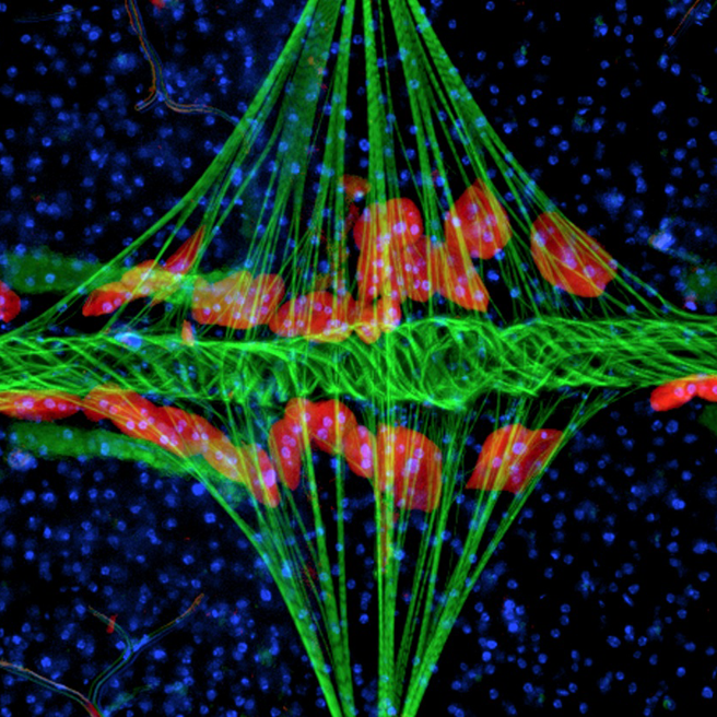
English: This fluorescence image details the structural organization of the heart of the mosquito Anopheles gambiae. The point of view is top down, with the mosquito's body lying horizontally with its head to the left (outside of the image). Muscle is labeled with phalloidin (green), and shows the tube-like heart extending horizontally across the body and the diamond shaped alary muscles projecting vertically onto the heart. The pericardial cells, labeled with 568 nm-Immunoglobulin G (red), are pinocytic cells that flank the heart. Cell nuclei are labeled with Hoechst 33342 (blue).
Image Credit: Jonas G. King and Julián F. Hillyer, Department of Biological Sciences, Vanderbilt University. Issue image, PLOS Pathogens, November 2012. |
| Date | |
| Source | King JG, Hillyer JF (2012) Infection-Induced Interaction between the Mosquito Circulatory and Immune Systems. PLoS Pathog 8(11): e1003058. doi:10.1371/journal.ppat.1003058 |
| Author | King JG, Hillyer JF |
Licensing
This file is licensed under the
Creative Commons
Attribution 2.5 Generic license.
- You are free:
- to share – to copy, distribute and transmit the work
- to remix – to adapt the work
- Under the following conditions:
- attribution – You must give appropriate credit, provide a link to the license, and indicate if changes were made. You may do so in any reasonable manner, but not in any way that suggests the licensor endorses you or your use.
File history
Click on a date/time to view the file as it appeared at that time.
| Date/Time | Thumbnail | Dimensions | User | Comment | |
|---|---|---|---|---|---|
| current | 20:13, 24 July 2015 |
| 656 × 656 (670 KB) | Cmdrjameson | Compressed with pngout. Reduced by 260kB (27% decrease). |
| 02:07, 2 December 2012 |
 | 656 × 656 (930 KB) | Daniel Mietchen | User created page with UploadWizard |
File usage
The following pages on the English Wikipedia use this file (pages on other projects are not listed):
Global file usage
The following other wikis use this file:
- Usage on ar.wikipedia.org
- Usage on bn.wikipedia.org
- Usage on hy.wikipedia.org
- Usage on ko.wikipedia.org
- Usage on outreach.wikimedia.org
- Usage on si.wikipedia.org