
Original file (700 × 743 pixels, file size: 82 KB, MIME type: image/jpeg)
| This is a file from the
Wikimedia Commons. Information from its
description page there is shown below. Commons is a freely licensed media file repository. You can help. |
Summary
| Description |
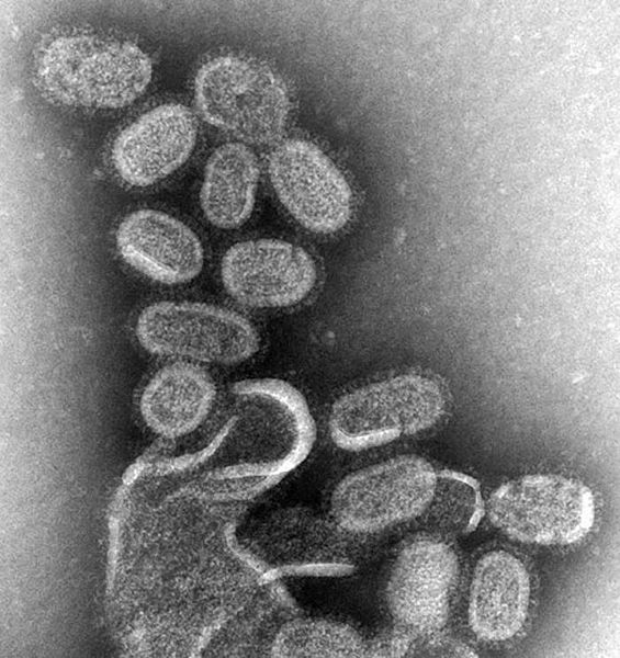
English: This negative stained transmission electron micrograph (TEM) shows recreated 1918 influenza virions that were collected from supernatants of 1918-infected Madin-Darby Canine Kidney (MDCK) cells cultures 18 hours after infection.
To separate these virions, the MDCK cells are spun down (centrifugation), and the 1918 virus in the fluid is immediately fixed for negative staining. The solid mass in lower center contains MDCK cell debris that did not spin down during the procedure. Dr. Terrence Tumpey, one of the organization’s staff microbiologists and a member of the National Center for Infectious Diseases (NCID), recreated the 1918 influenza virus in order to identify the characteristics that made this organism such a deadly pathogen. Research efforts such as this, enables researchers to develop new vaccines and treatments for future pandemic influenza viruses. The 1918 Spanish flu epidemic was caused by an influenza A (H1N1) virus, killing more than 500,000 people in the United States, and up to 50 million worldwide. The possible source was a newly emerged virus from a swine or an avian host of a mutated H1N1 virus. Many people died within the first few days after infection, and others died of complications later. Nearly half of those who died were young, healthy adults. Influenza A (H1N1) viruses still circulate today after being introduced again into the human population in the 1970s.Ελληνικά: EM of influenza virus.jpg.
Tiếng Việt: siêu vi cúm qua hiển vi điện tử. |
||
| Date | |||
| Source |
|
||
| Author |
|
||
| Permission ( Reusing this file) |
PD-USGov-HHS-CDC English: None - This image is in the public domain and thus free of any copyright restrictions. As a matter of courtesy we request that the content provider be credited and notified in any public or private usage of this image. |
Licensing
This image is a work of the
Centers for Disease Control and Prevention, part of the
United States Department of Health and Human Services, taken or made as part of an employee's official duties. As a work of the
U.S. federal government, the image is in the
public domain.
|
Original upload log
(All user names refer to en.wikipedia)
- 2006-10-26 03:31 TimVickers 700×743×8 (83774 bytes) CDC, CDC Public Health Image Library (PHIL), http://phil.cdc.gov/Phil/details.asp
File history
Click on a date/time to view the file as it appeared at that time.
| Date/Time | Thumbnail | Dimensions | User | Comment | |
|---|---|---|---|---|---|
| current | 13:41, 10 August 2007 |
| 700 × 743 (82 KB) | ToNToNi | {{Information |Description=CDC, CDC Public Health Image Library (PHIL), http://phil.cdc.gov/Phil/details.asp |Source=Originally from [http://en.wikipedia.org en.wikipedia]; description page is/was [http://en.wikipedia.org/?title=Image%3AEM_of_i |
File usage
- Emergent virus
- Influenza
- Influenza A virus
- User:JenOttawa/Notes/practice
- User:Mr. Ibrahem/Influenza
- Wikipedia:Today's featured article/January 1, 2007
- Wikipedia:Today's featured article/January 2007
- Wikipedia:VideoWiki/Influenza
- Portal:Medicine/Selected Article
- Portal:Medicine/Selected Article/10
- Portal:Medicine/Selected Article Archive
- Portal:Medicine/Selected article/8, 2008
Global file usage
The following other wikis use this file:
- Usage on af.wikipedia.org
- Usage on an.wikipedia.org
- Usage on ar.wikipedia.org
- Usage on as.wikipedia.org
- Usage on awa.wikipedia.org
- Usage on azb.wikipedia.org
- Usage on az.wikipedia.org
- Usage on bat-smg.wikipedia.org
- Usage on ba.wikipedia.org
- Usage on be-tarask.wikipedia.org
- Usage on be.wikipedia.org
- Usage on bg.wikipedia.org
- Usage on bn.wikipedia.org
- Usage on bo.wikipedia.org
- Usage on br.wikipedia.org
- Usage on bs.wikipedia.org
- Usage on bxr.wikipedia.org
- Usage on ca.wikipedia.org
- Usage on cdo.wikipedia.org
- Usage on ckb.wikipedia.org
- Usage on csb.wikipedia.org
- Usage on cs.wikipedia.org
- Usage on da.wikipedia.org
- Usage on de.wikipedia.org
- Usage on en.wikibooks.org
- Usage on en.wikinews.org
- Usage on et.wikipedia.org
- Usage on eu.wikipedia.org
- Usage on fa.wikipedia.org
- Usage on fi.wikipedia.org
- Usage on fj.wikipedia.org
- Usage on fr.wikipedia.org
- Usage on fr.wikiversity.org
- Usage on ga.wikipedia.org
- Usage on hak.wikipedia.org
- Usage on hr.wikipedia.org
- Usage on hy.wikipedia.org
- Usage on ia.wikipedia.org
View more global usage of this file.