
Size of this preview:
800 × 532 pixels. Other resolutions:
320 × 213 pixels |
640 × 426 pixels |
1,024 × 682 pixels |
1,280 × 852 pixels |
1,809 × 1,204 pixels.
Original file (1,809 × 1,204 pixels, file size: 808 KB, MIME type: image/jpeg)
| This is a file from the
Wikimedia Commons. Information from its
description page there is shown below. Commons is a freely licensed media file repository. You can help. |
| Description |
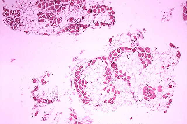
Deutsch: Histopathologisches Bild eines Querschnitts aus dem Wadenmuskel (Muskulus gastrocnemius) eines Patienten, der an Muskeldystrophie Typ Duchenne verstarb. Das Bild verdeutlicht, in welchem Ausmaß die (rot gefärbten) Muskelfasern durch Fettzellen (optisch leer = weiß) ersetzt wurden.
English: Histopathology of gastrocnemius muscle from patient who died of pseudohypertrophic muscular dystrophy, Duchenne type. Cross section of muscle shows extensive replacement of muscle fibers by adipose cells. |
||
| Date | |||
| Source |
http://phil.cdc.gov/phil/home.asp ID#: 70 US Department of Health and Human Services
|
||
| Author | Dr. Edwin P. Ewing, Jr. | ||
| Permission ( Reusing this file) |
PD-USGov-HHS-CDC English: None - This image is in the public domain and thus free of any copyright restrictions. As a matter of courtesy we request that the content provider be credited and notified in any public or private usage of this image. |
This image is a work of the
Centers for Disease Control and Prevention, part of the
United States Department of Health and Human Services, taken or made as part of an employee's official duties. As a work of the
U.S. federal government, the image is in the
public domain.
|
File history
Click on a date/time to view the file as it appeared at that time.
| Date/Time | Thumbnail | Dimensions | User | Comment | |
|---|---|---|---|---|---|
| current | 00:46, 3 December 2013 |
| 1,809 × 1,204 (808 KB) | Splintercellguy | Upload higher-resolution version |
| 10:33, 13 August 2006 |
 | 700 × 465 (50 KB) | Der Lange | {{Information| |Description= * '''de:''' Histopathologisches Bild eines Querschnitts aus dem Wadenmuskel (Muskulus gastrocnemius) eines Patienten, der an Muskeldystrophie Typ Duchenne verstarb. Das Bild verdeutlicht, in welchem Ausmaß die (rot gefärbten |
File usage
The following pages on the English Wikipedia use this file (pages on other projects are not listed):
Global file usage
The following other wikis use this file:
- Usage on ar.wikipedia.org
- Usage on be.wikipedia.org
- Usage on bs.wikipedia.org
- Usage on ca.wikipedia.org
- Usage on cs.wikipedia.org
- Usage on de.wikipedia.org
- Usage on de.wikibooks.org
- Usage on el.wikipedia.org
- Usage on es.wikipedia.org
- Usage on et.wikipedia.org
- Usage on eu.wikipedia.org
- Usage on fa.wikipedia.org
- Usage on fi.wikipedia.org
- Usage on gl.wikipedia.org
- Usage on he.wikipedia.org
- Usage on hy.wikipedia.org
- Usage on id.wikipedia.org
- Usage on ja.wikipedia.org
- Usage on la.wikibooks.org
- Usage on mk.wikipedia.org
- Usage on no.wikipedia.org
- Usage on or.wikipedia.org
- Usage on pl.wikipedia.org
- Usage on ru.wikipedia.org
- Usage on sh.wikipedia.org
- Usage on sr.wikipedia.org
- Usage on sv.wikipedia.org
- Usage on ta.wikipedia.org
- Usage on th.wikipedia.org
- Usage on tl.wikipedia.org
- Usage on tr.wikipedia.org
- Usage on uk.wikipedia.org
- Usage on vi.wikipedia.org
- Usage on www.wikidata.org
- Usage on zh.wikipedia.org