Size of this preview:
337 × 599 pixels. Other resolutions:
135 × 240 pixels |
270 × 480 pixels |
432 × 768 pixels |
576 × 1,024 pixels |
1,650 × 2,933 pixels.
Original file (1,650 × 2,933 pixels, file size: 1.27 MB, MIME type: image/jpeg)
| This is a file from the
Wikimedia Commons. Information from its
description page there is shown below. Commons is a freely licensed media file repository. You can help. |
Summary
| Description |
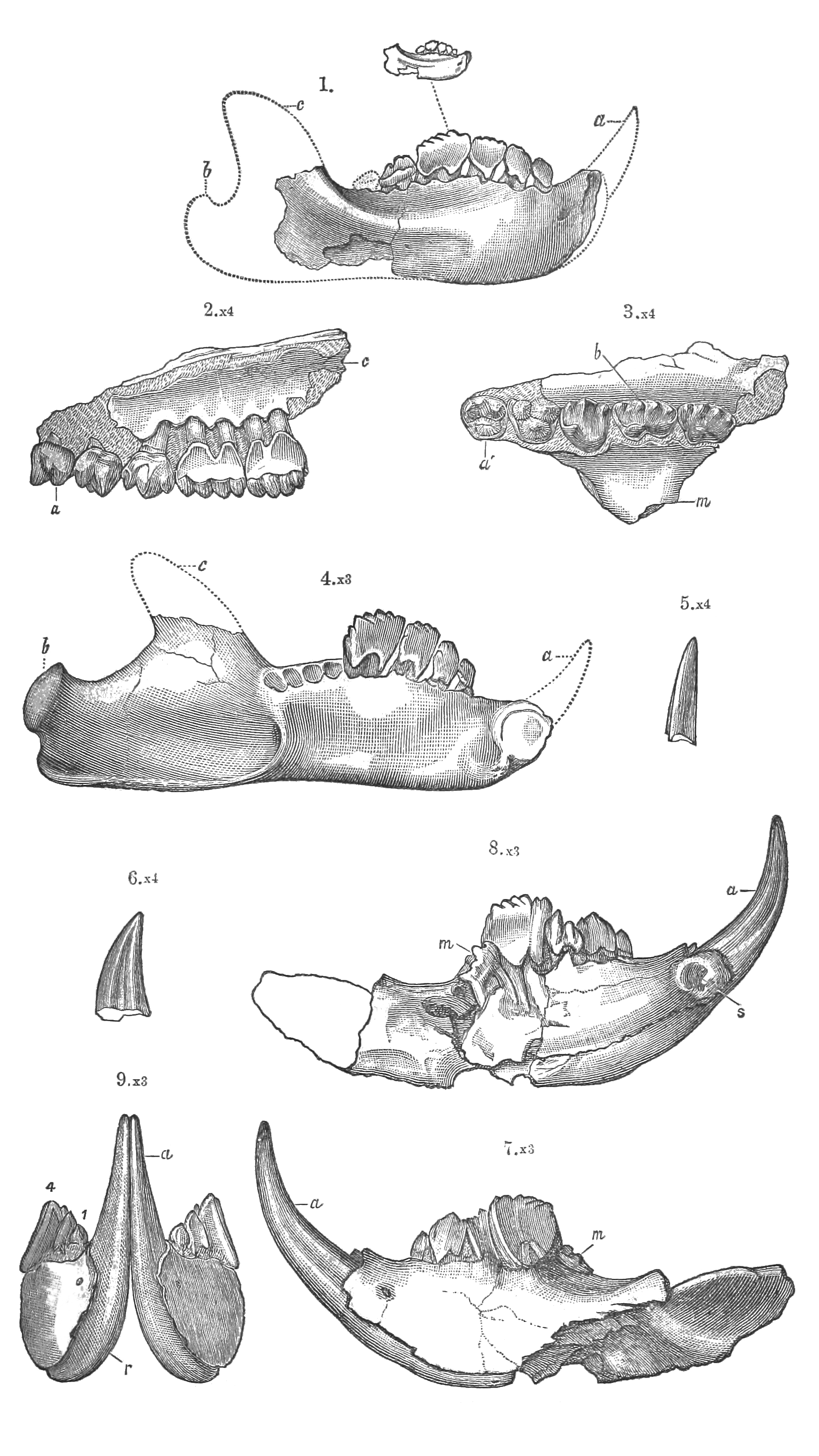
English: PLATE VII.
In Figures 2 and 3, a’, first premolar; b, fourth premolar; c, second molar ; m, malar arch. In the lower jaws, a, incisor ; b, condyle ; c, coronoid process ; m, molar ; r, root of incisor ; s, symphyseal surface. |
| Date | |
| Source | Marsh, O. C. (1887). I.— American Jurassic Mammals. Geological Magazine (Decade III), 4(07), 289-299. |
| Author | Othniel Charles Marsh |
Licensing
|
This work is in the public domain in its country of origin and other countries and areas where the copyright term is the author's life plus 70 years or fewer. This work is in the public domain in the United States because it was published (or registered with the U.S. Copyright Office) before January 1, 1929. | |
| This file has been identified as being free of known restrictions under copyright law, including all related and neighboring rights. | |
File history
Click on a date/time to view the file as it appeared at that time.
| Date/Time | Thumbnail | Dimensions | User | Comment | |
|---|---|---|---|---|---|
| current | 19:23, 4 April 2015 |
| 1,650 × 2,933 (1.27 MB) | Animalparty | BW, cleared, cropped |
| 21:28, 2 November 2014 |
 | 1,822 × 3,170 (3.37 MB) | Animalparty | User created page with UploadWizard |
File usage
The following pages on the English Wikipedia use this file (pages on other projects are not listed):
Global file usage
The following other wikis use this file:
- Usage on ca.wikipedia.org
- Usage on en.wiktionary.org
- Usage on es.wikipedia.org
- Usage on eu.wikipedia.org
- Usage on fa.wikipedia.org
- Usage on hu.wikipedia.org
- Usage on it.wikipedia.org
- Usage on species.wikimedia.org
- Usage on www.wikidata.org