
Original file (2,048 × 1,532 pixels, file size: 912 KB, MIME type: image/jpeg)
| This is a file from the
Wikimedia Commons. Information from its
description page there is shown below. Commons is a freely licensed media file repository. You can help. |
Summary
| Description |
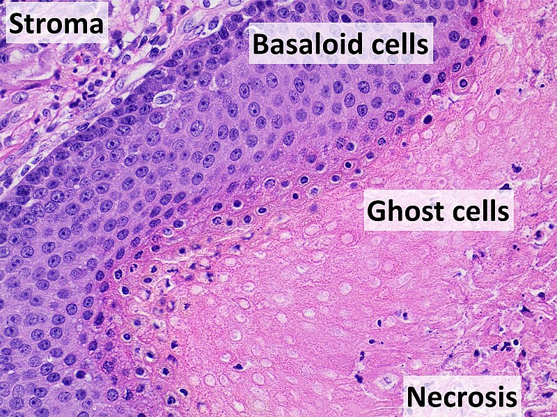
English: Histopathology of pilomatricoma, high magnification, H&E stain, showing the characteristic components: Reference
Gallery of the same case
|
| Date | |
| Source | Own work |
| Author |
.jpg) - Reusing images - Conflicts of interest: None Consent note: Consent from the patient or patient's relatives is regarded as redundant, because of absence of identifiable features ( List of HIPAA identifiers) in the media and case information ( See also HIPAA case reports guidance). |


Licensing
| This file is made available under the Creative Commons CC0 1.0 Universal Public Domain Dedication. | |
| The person who associated a work with this deed has dedicated the work to the
public domain by waiving all of their rights to the work worldwide under copyright law, including all related and neighboring rights, to the extent allowed by law. You can copy, modify, distribute and perform the work, even for commercial purposes, all without asking permission.
|
File history
Click on a date/time to view the file as it appeared at that time.
| Date/Time | Thumbnail | Dimensions | User | Comment | |
|---|---|---|---|---|---|
| current | 14:43, 25 September 2021 |
| 2,048 × 1,532 (912 KB) | Mikael Häggström | Uploaded a work by {{Mikael Häggström|cat=Micrographs of the skin|consent=noid}} from {{Own}} with UploadWizard |