
Size of this preview:
604 × 600 pixels. Other resolutions:
242 × 240 pixels |
483 × 480 pixels |
902 × 896 pixels.
Original file (902 × 896 pixels, file size: 386 KB, MIME type: image/jpeg)
| This is a file from the
Wikimedia Commons. Information from its
description page there is shown below. Commons is a freely licensed media file repository. You can help. |
Summary
| Description |
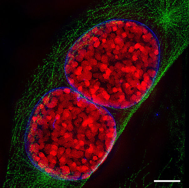
English: Light-optical section through two mouse cell nuclei in prophase, recorded with 3D Structured Illumination Microscopy (3D-SIM-microscopy). condensed chromosomes are red, the nuclear envelope blue and microtubuli, which belong to the cytoskeleton, are green. Scale bar is 5 µm.
For further information see: Schermelleh L, Carlton PM, Haase S, Shao L, Winoto L, Kner P, Burke B, Cardoso MC, Agard DA, Gustafsson MG, Leonhardt H, Sedat JW (June 2008). "Subdiffraction multicolor imaging of the nuclear periphery with 3D structured illumination microscopy". Science (journal) 320 (5881): 1332–6.
DOI:
10.1126/science.1156947.
PMID
18535242.
Deutsch: Lichtoptischer Schnitt durch zwei Mauszellkerne in der Prophase aufgenommen mit 3D-SIM. Die kondensierten Chromosomen sind rot, die Kernhülle blau und Mikrotubuli, ein Bestandteil des Zellskeletts, grün eingefärbt. Der Maßstab entspricht 5 µm. Für weitere informationen siehe die oben angegebene Veröffentlichung. |
| Date | |
| Source | Lothar Schermelleh |
| Author | Lothar Schermelleh |
| Permission ( Reusing this file) |
This file is licensed under the
Creative Commons
Attribution-Share Alike 3.0 Unported license.
|
3D-SIM images


File history
Click on a date/time to view the file as it appeared at that time.
| Date/Time | Thumbnail | Dimensions | User | Comment | |
|---|---|---|---|---|---|
| current | 18:41, 16 January 2009 |
| 902 × 896 (386 KB) | Dietzel65 | == Beschreibung == {{Information |Description={{en|1=(to be added soon) For further information see: {{cite journal |author=Schermelleh L, Carlton PM, Haase S, Shao L, Winoto L, Kner P, Burke B, Cardoso MC, Agard DA, Gustafsson MG, Leonhardt H, Sedat JW |
File usage
The following pages on the English Wikipedia use this file (pages on other projects are not listed):
Global file usage
The following other wikis use this file:
- Usage on bn.wikipedia.org
- Usage on bs.wikipedia.org
- Usage on ca.wikipedia.org
- Usage on de.wikipedia.org
- Usage on el.wikipedia.org
- Usage on es.wikipedia.org
- Usage on eu.wikipedia.org
- Usage on id.wikipedia.org
- Usage on ja.wikipedia.org
- Usage on nl.wikipedia.org
- Usage on ru.wikipedia.org
- Usage on sh.wikipedia.org
- Usage on si.wikipedia.org
- Usage on sl.wikipedia.org
- Usage on sq.wikipedia.org
- Usage on sr.wikipedia.org
- Usage on ta.wikipedia.org
- Usage on uk.wikipedia.org
- Usage on uz.wikipedia.org
- Usage on vi.wikipedia.org
- Usage on zh.wikipedia.org